





Despite the fact that we quite literally embody our own bodies, they can still be a source of horror in the right (or wrong) circumstances. It’s one of the reasons roughly 4% of US medical students drop out, not due to poor grades but the realization that this maybe isn’t for them.
We’ve gathered some unsettling, creepy but perhaps still interesting pictures about the unusual things that can go on in and outside of the human body. Be warned, some of these images can be unpleasant. Otherwise, settle in as you scroll through, upvote your favorites and be sure to share your thoughts in the comments down below.
#41 A Giant Gastric Trichobezoar (Hair Ball)! A Bezoar Is A Tightly Packed Collection Of Undigested Material That Is Unable To Exit The Stomach, Most Being Hair-Trichobezoars
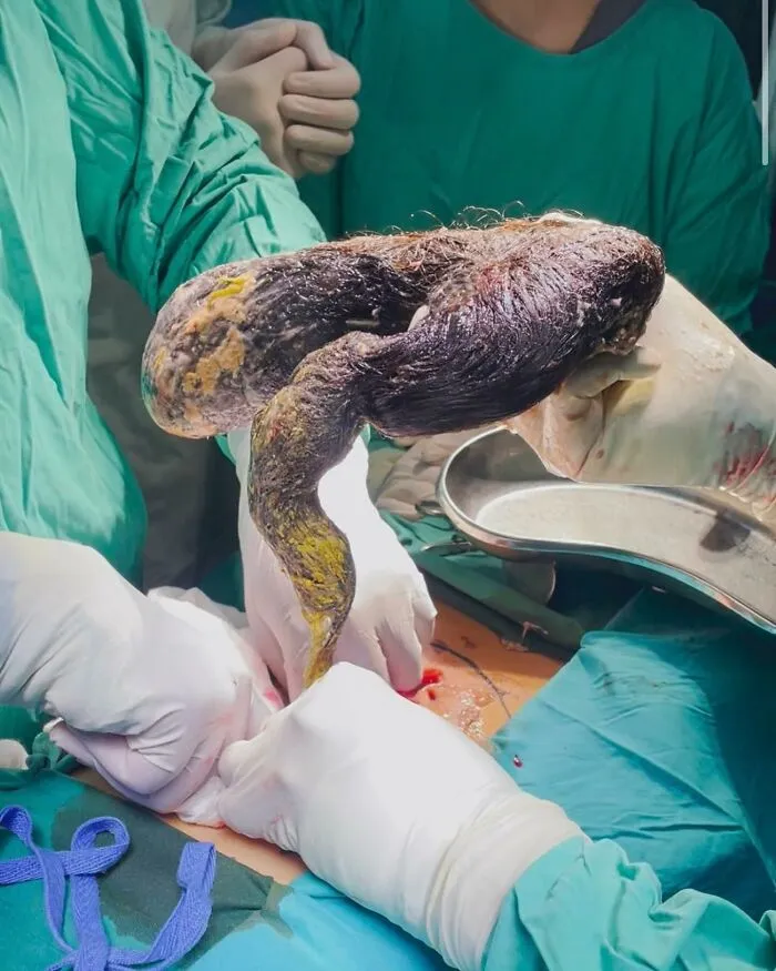
This 16-year-old female with a positive psychiatric history of OCD had uncontrollable repetitive thoughts with urge to pull (trichotillomania) and eat hair (trichophagia).
She presented with epigastric pain for the past six months with progressive loss of apetite and early satiety. Vomiting with increasing frequency for the last two months. Lean patient with hair cropped short.
On examination, soft, non-tender abdomen with no distention. Firm to hard mass palpable, craniocaudally extending from the below the subcostal margins to the infraumbilical region and horizontally between both midclavicular lines.
Mass was mobile and moved with respiration. No other positive findings on physical examination.
XRay Abdomen showed a radio-opaque shadow occupying most of the abdominal cavity. CECT showed a large intraluminal mass in the stomach extending up to the
second part of duodenum consistent with a gastric bezoar. Laparotomy and extraction of trichobezoar done successfully. The bezoar had formed into a cast of the stomach and the first and second parts of the duodenum. Uneventful post-op course. Patient discharged over to psychiatric care for further management.
Report
10points
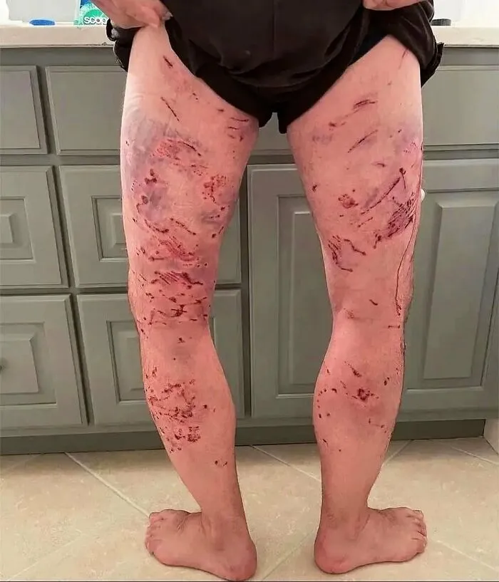
#42 Consequence Of Multiple Dog Bites! Bites And Maulings By Dogs, Sometimes Fatal, Are A Worldwide Problem And Particularly Affect Children
Report
9points
#43 More Than 200 Bladder Stones Found In An 81-Year-Old Man
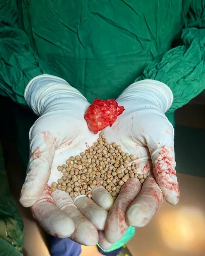
The patient in this case is an elderly man who has had urinary stasis obstruction for a long time due to benign prostate hyperplasia (BPH).
Due to urinary stasis, numerous stones have formed inside the bladder.
Given the large number of bladder stones and the patient’s symptoms, the management plan involved a multidisciplinary approach, including Transurethral resection of the prostate (TURP): To alleviate bladder outlet obstruction caused by BPH and reduce the risk of recurrent stone formation, an open bladder stone removal, which led to the removal of a mind-blowing number of over 200 stones!
Bladder stones are solid calculi that are primarily found in the urinary bladder. While often calcified, they may also be composed of non-calcific material.
Urinary stasis, such as BPH or neurogenic bladder disorder, is the primary cause. Most such stones are newly formed in the bladder. Some come from the kidneys either as a stone or a sloughed papilla.
Any foreign body left in the bladder that is not spontaneously expelled will eventually form layers of stone material and develop into calculus. In 50% of cases the stone composition is uric acid.
In regards to surgical therapy, endoscopic surgery is used.
Cystolitholapaxy (to break up bladder stones into smaller pieces and remove them), can be achieved by disruptive or ablative therapy, including lasers, pneumatic-powered mechanical contact jackhammer, ultrasound, and direct mechanical crushing with a lithotrite.
Extracorporeal shockwave lithotripsy is also used.
In some cases of extremely large bladder stones or prostates, open suprapubic surgery is done. This allows for the removal of the intact stone, followed by an open prostatectomy (for prostates more than 75 g in size).
The advantage of an open suprapubic cystostomy for bladder stone removal is the reduced surgery time (half the time compared to endoscopic means), easy removal of large or multiple stones, ability to remove stones that might be difficult or hard to fragment with endoscopic treatments and the ability to handle stones that are stuck to the bladder lining.
Report
9points
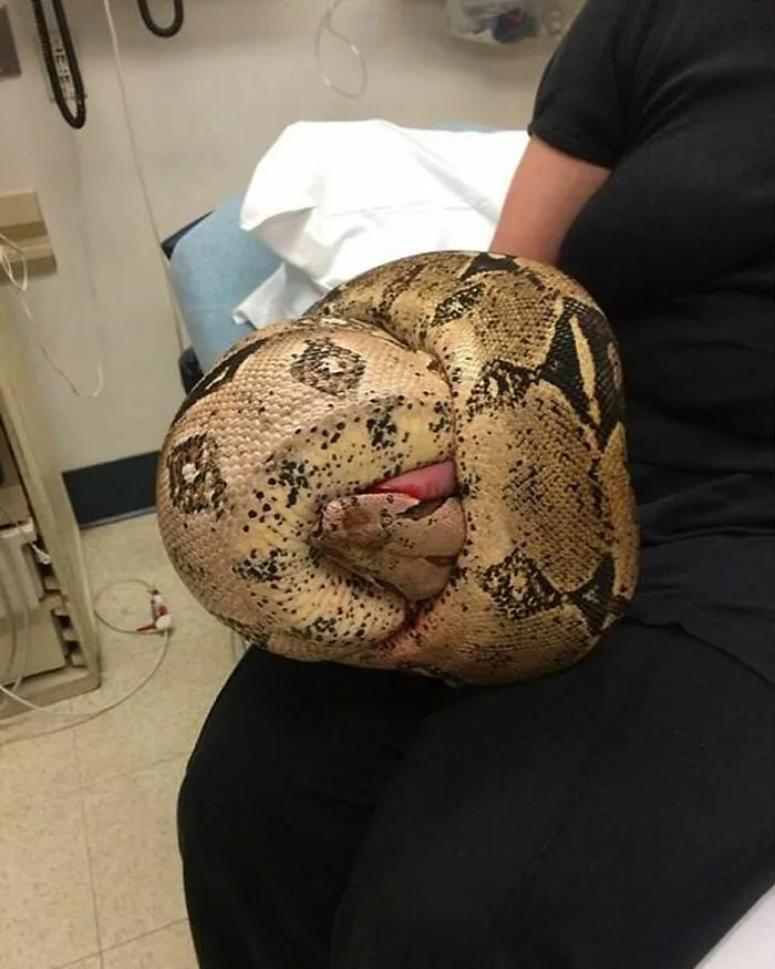
#44 You Arrive On Scene Of A Residential Home And Find A Pet Snake Constricting Around Someone’s Hand
Report
9points
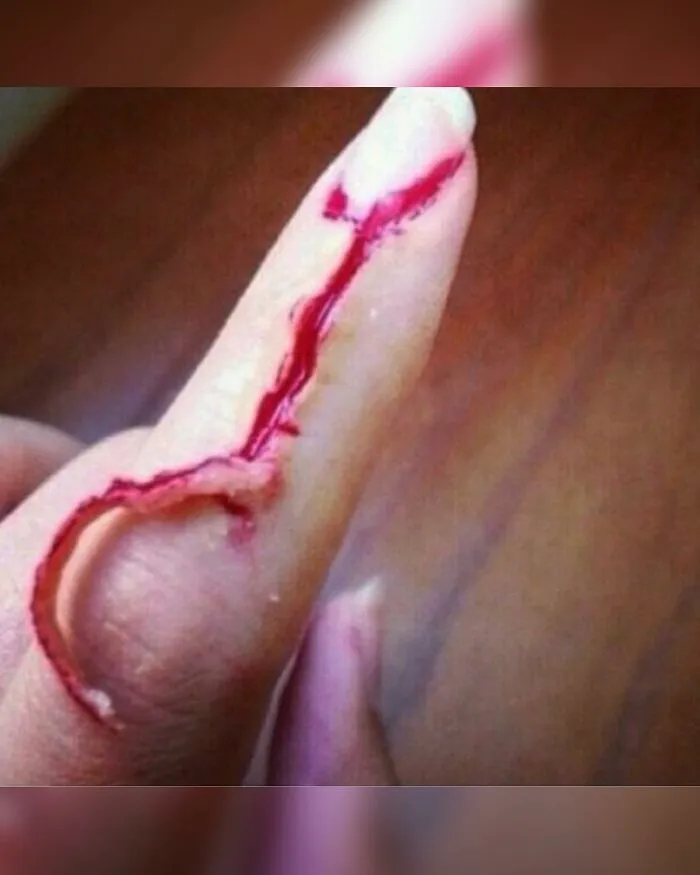
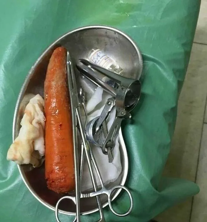
#46 But With A Sprinkle Of Imagination We Can All Assume What Happened Here. This Was Extracted As A Whole, Let’s Just Agree That Working In A Hospital Isn't For The Faint Hearted
Report
9points
#47 Degloving Injury After Hand Got Caught At A Factory Machine!! Literally Came Off Like A Glove
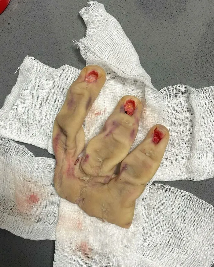
This 38-year-old male patient presented to the emergency department after getting his hand crushed and degloved in a heavy equipment machine at the factory he works.
Notice the extensive section of skin that got completely torn off the underlying tissue, severing its blood supply. De-gloving injury is named by analogy to the process of removing a glove. As simple as it sounds.
The patient underwent emergency surgery, and replantation was performed with the assistance of a microscopic magnification.
Formal replantation (re-attachment) involves debridement (removal of dead or contaminated tissues), repair of the underlying tendons and muscles if injured, and revascularization of the degloved skins, or when these are not possible, skin grafts or skin flaps.
This requires VERY delicate instruments and working hands.
Due to poor blood circulation and extent of the trauma the vasculature, the middle and distal phalanges of the index finger were amputated.
Report
9points
#48 Screw To The Eye! Ouch. An Unfortunate Accident Of A 3 Year Old Boy With A Screw Penetrating His Left Eye After Lawnmower Spits Out Bolt!!
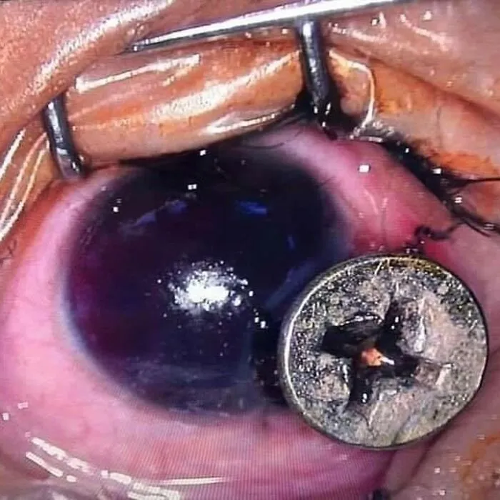
His dad was using the petrol-powered tool which was slung over his shoulder when the accident happened.
As he mowed the grass, the blade struck a one-inch long metal screw hidden in the lawn.
The piece of metal flicked into the air and flew straight into the toddler’s eyeball.
Doctors found that miraculously the screw missed the cornea and retina. X-rays also reveal just how deep the piece of metal became lodged into his eye socket - almost reaching the skull.
Doctors performed successful surgery to remove the screw.
The ophthalmologist that did the operation said that they will check the wound over the coming days but they are confident the boy will be able to see again.
He said: “Fortunately, the screw struck his eyeball but did not go into the lens, which thankfully has spared his sight.
“However, due to the accident his eye bled badly and now is still bloodshot so there will be follow up treatment to help the boy see again.
‘’We’re pleased with the outcome of the surgery. If the screw had been slightly to the left he would have been blinded.’’
Report
9points
#49 A 20-Year-Old Man Was Subjected To High-Voltage Electric Burn
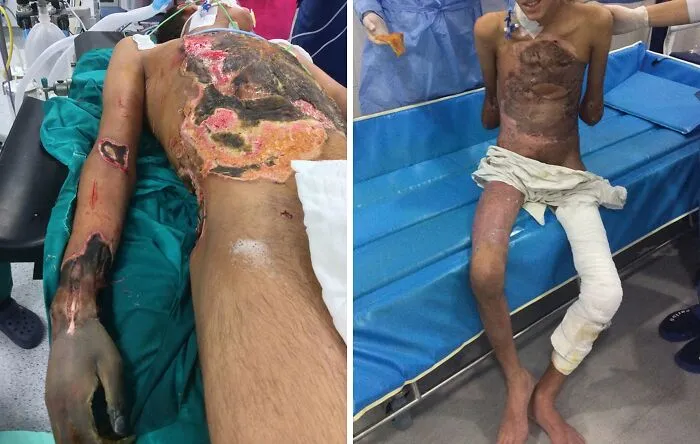
The patient was admitted to a military medical complex 12 days after the accident had happened. The patient had a 40% total body surface area burn involving the upper limbs, anterior and posterior trunks, and the left thigh (third- and fourth-degree burns).
The first photo shows the patient on table just before the first operation (mummified right hand and distal forearm, anterior trunk, and lower posterior trunk show third- and fourth-degree burns with severed tissues.
The aim was to stabilize the patient by conducting lifesaving operations in multiple scheduled sessions, bilateral below-elbow amputations, escharotomies, and excision of affected ribs and cartilages. A left latissimus dorsi flap was used to cover the left side of the anterior chest wall. Skin grafting (split thickness) was done to cover the raw areas. Multiple aggressive operations by a multidisciplinary team saved the patient’s life.
Last photo shows the patient just before being discharged and with wounds totally healed.
Report
9points
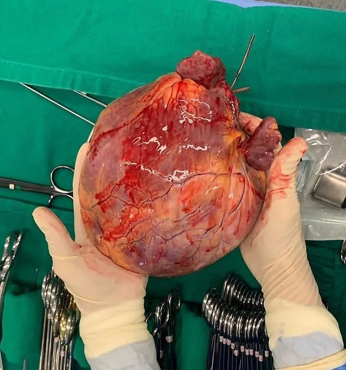
#51 A Mega-Size Globular Heart!! This Very Large Heart Has A Globoid Shape Because All Of The Chambers Are Dilated. It Felt Very Flabby, And The Myocardium Was Poorly Contractile
Report
9points
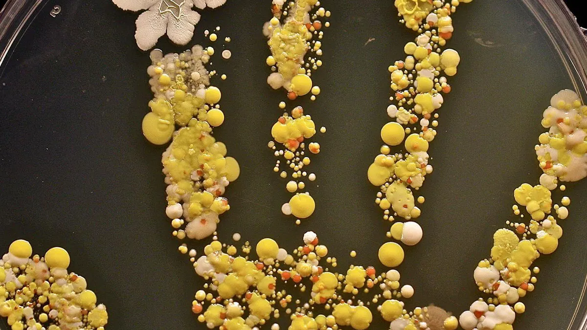
#52 Dinner Is Served — Petri Dish With A Colony Of Bacteria On A Hand!! This Is The Handprint Of An 8-Year-Old Boy Covered In Bacteria
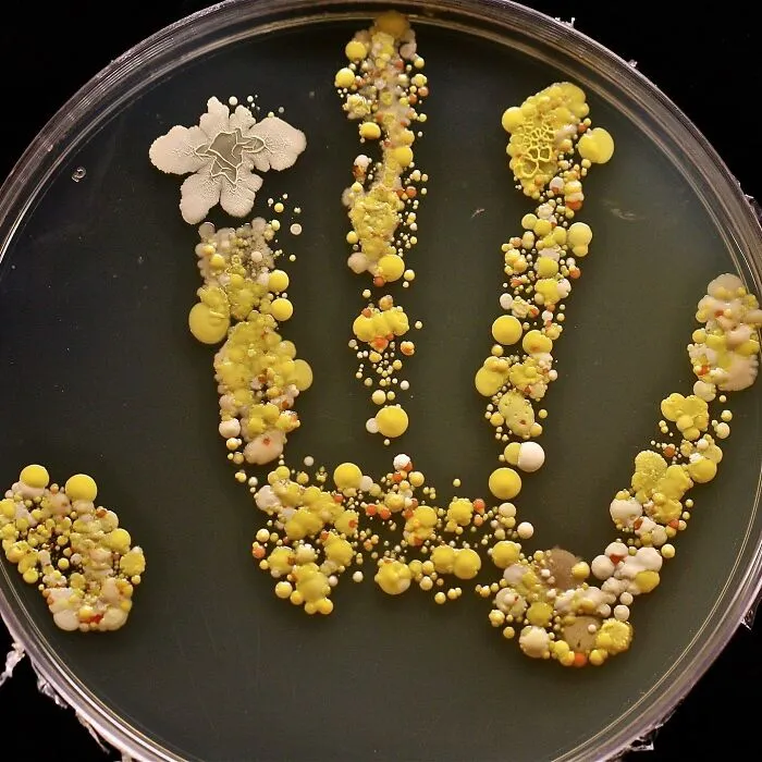
The boy came in from playing outside, and his mom decided to put his hand print inside a large Petri dish, incubated it for two days, and ended up with a colorful germ garden!
Consider for a moment what you've touched today. After all, our hands do all sorts of things for us – they open doors, handle money at the wet market, hold on to poles on public buses and trains, all the time while grasping our phones. They often also act as the barrier between our coughs and the rest of the world.
Up to 80% of all infections are transmitted by hands.
The colony on the hand would consist of various bacteria, potentially including both gram-positive and gram-negative species. These bacteria could belong to different genera and species, such as Staphylococcus, Streptococcus, or Escherichia coli, among others. The colony may exhibit different morphological characteristics, such as size, shape, and coloration, depending on the specific bacteria present.
Additionally, the colony might demonstrate characteristics indicative of bacterial growth, such as a visible texture or elevation. It could display features like smoothness, roughness, or irregularity. The Petri dish would provide a suitable environment for these bacteria to multiply, as it would contain a culture medium that supports their growth and sustenance.
Report
8points
#53 Avocado Slicing Injuries Are On The Rise Due To Lack Of Handling Skills
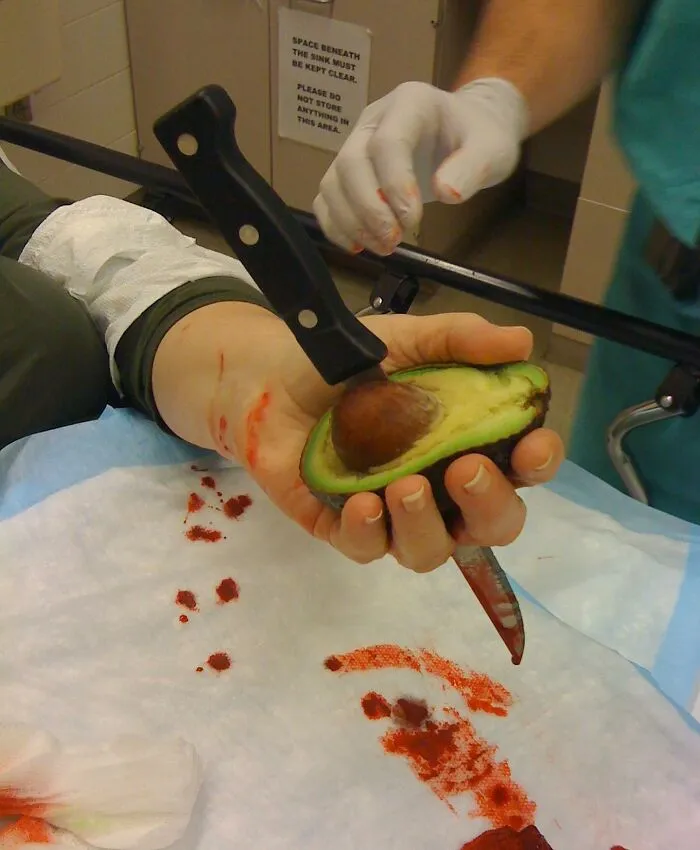
Avocado slicing injuries are on the rise due to lack of handling skills. This patient accidentally stabbed her hand while trying to cut an avocado and remove the pit. Guess it’s simply a matter of the knife slipping.
The patient used the knife to split an avocado in half and then tried to get the stone out with a spoon but with no luck. She put the knife in to the pip and turned it clock wise to prise it out but had no luck, so she put the knife under the pip to dig it out and pushed to far.
Avocado is one of the most popular brunch items - but the task of removing the pit due to lack of handling skills is proving to be a real kitchen hazard.
Classically, patients hold the avocado in their non-dominant hand while using a knife to cut/peel the fruit with their dominant hand.
The mechanism of injury is usually a stabbing injury to the non-dominant hand as the knife slips past the stone, through the soft fruit.
These stabbing injuries have the potential to cause significant neurovascular, tendinous and/or bony injuries.
Report
8points
#54 A 15 Year Old Male With A Foreign Body - Wounded By A Wood Splinter
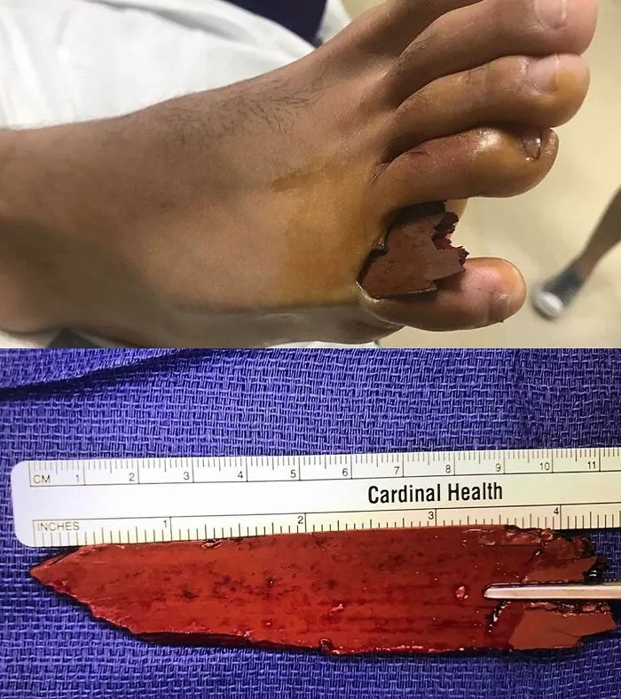
An impalement injury is a traumatic condition characterized by the penetration of a foreign object into the body.
In the specific case mentioned, a segment of wood penetrated the soft tissues of the right foot, specifically entering the interdigital space between the fourth and fifth toes.
This patient presented to the emergency department with an 11cm long piece of wood embedded in his right foot.
He was playing on a dock of a lake in Miami, when he tried jumping into the water, the wood sticking out of the dock penetrated the skin entering between the right foot's fourth toe and the pinkie toe area.
Luckily, there was no damage to nerves, ligaments or vessels. The object was found just under the skin and was surgically pulled out in one piece with the foot being under local anesthetics.
Report
8points
#55 Always Careful When Stepping. This Patient Stumbled And Accidentally Stepped On A Cuticle Clipper!! You Don’t Have To Be Sharp To Know That This One Hurt
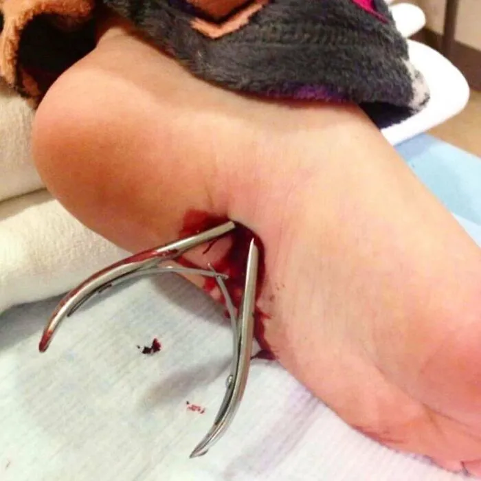
While walking barefoot in the shower room, this patient got distracted for just a second, and the next thing he knows this clipper went right through his foot with its top and sharp edge lodged into the plantar aspect or sole of his left foot.
Luckily, he had surgery and recovered totally, but not sure he’ll enjoy the showering again.
Luckily, he had surgery and recovered totally, but not sure he’ll enjoy the showering again.
Report
8points
#56 This Case Involves A Malfunctioning Piece Of Machinery That Shot A Metal Washer Into A Workers Eye
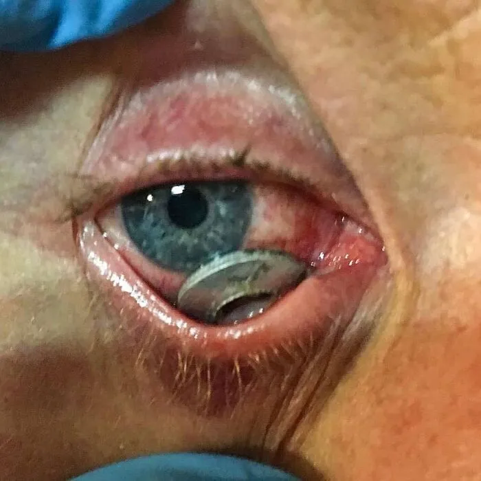
Unfortunately he was not wearing any eye protection or safety glasses at the time of the incident. Luckily the man was taken to a hospital and the foreign body was safely removed. He did not have any long term effects or vision loss. Even though this man was extremely lucky to have no lasting damage, vision loss and eye injury are very common with Intraocular injuries. Always remember to protect your eyes wear safety glasses!
Report
8points
#57 Plica Fimbriata Refers To The Small Folds In The Membrane On The Underside Of Your Tongue
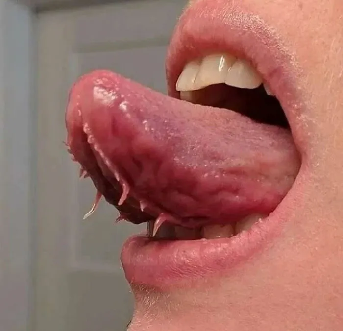
The folds tend to run parallel to, and on either side of your frenulum. The frenulum is the web of tissue that connects your tongue to the bottom of your mouth. These growths are harmless. They are normal residual tissue not completely reabsorbed by the body during the development and growth of the tongue.
Sometimes they can get caught in your teeth. If one of these bumps comes off, it can leave an open sore that’s vulnerable to infection. For most infections you may just need to use an antieptic mouthwash for a few days to keep the area clean.
Sometimes they can get caught in your teeth. If one of these bumps comes off, it can leave an open sore that’s vulnerable to infection. For most infections you may just need to use an antieptic mouthwash for a few days to keep the area clean.
Report
8points
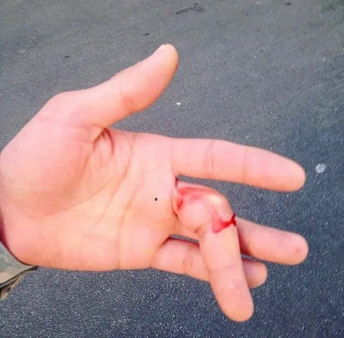
#58 Finger Was Forced Into Lateral Deviation And Dislocated Acutely During An Expedition
Report
7points
#59 Compartment Syndrome From Snake Bite
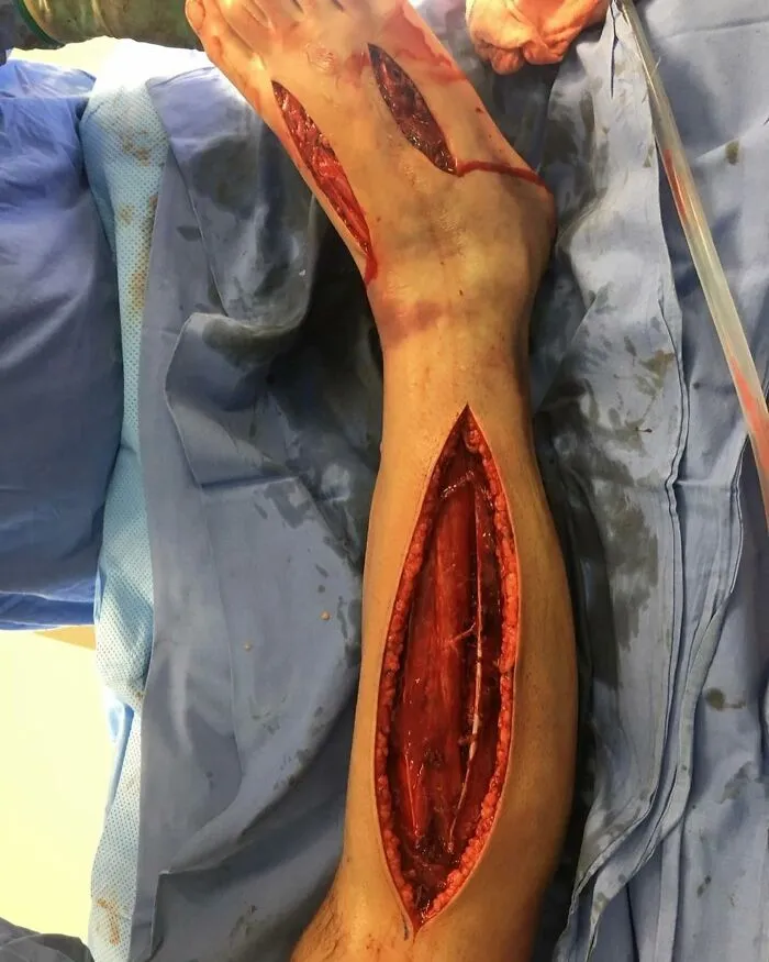
Snake bites can lead to various complications, one of the most severe being compartment syndrome. This condition occurs when increased pressure within a muscle compartment compromises blood flow, leading to tissue damage and potentially permanent disability if not promptly treated.
Compartment syndrome is a painful condition caused by increased intracompartmental pressure, compromising perfusion and resulting in muscle and nerve damage within that compartment, resulting in edema of the muscle. Because many muscles are encased in an inelastic, fascial compartment, edema increases the volume, and ultimately the pressure, within the compartment. As the pressure builds up, muscle perfusion decreases, resulting in further ischemic injury, which worsens the edema.
Compartment syndrome following a snake bite is a result of venom toxicity and tissue swelling. When a snake injects venom into its victim, the toxins can cause significant local tissue damage, inflammation, and swelling. This swelling within the confined space of a muscle compartment can quickly elevate pressure, impeding blood flow and depriving tissues of oxygen and nutrients.
Symptoms include severe pain that worsens with movement, swelling, numbness or tingling, and tightness or hardness of the affected area. In the case of a snake bite, these symptoms may develop rapidly and require immediate medical attention.
Early recognition and intervention are crucial in managing compartment syndrome. Treatment involves measures to relieve pressure within the affected compartment, such as fasciotomy—a surgical procedure to release the tight fascial compartments and relieve pressure.
It involves making incisions in the fascia surrounding the affected muscles to release built-up pressure, allowing blood flow to return to normal and preventing tissue damage.
Report
7points
#60 Here Is A Great Shot Showing The Brain With Its Covering Layer
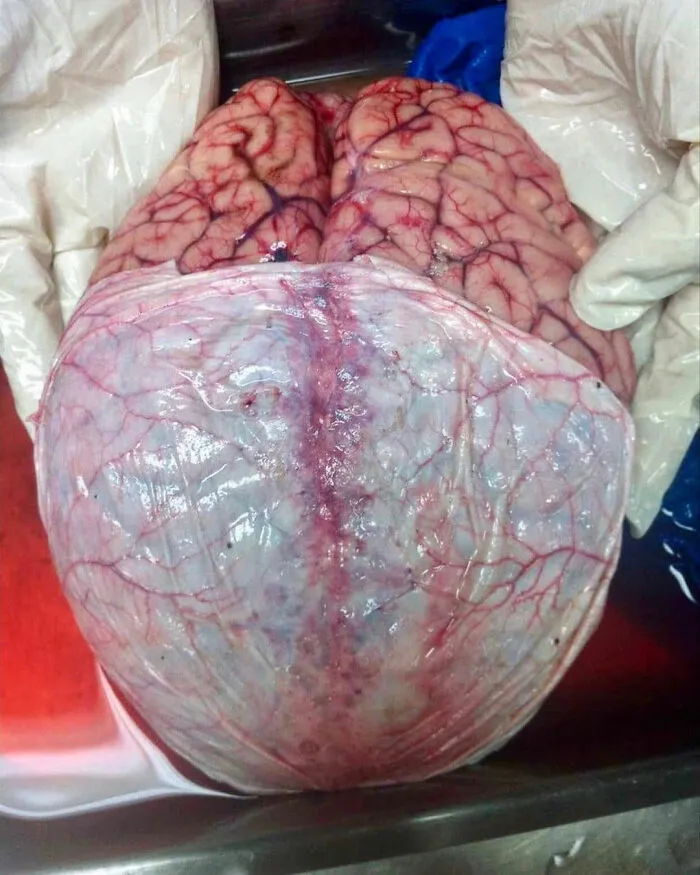
Below the skull and around the spinel cord are three special coverings called the meninges. You may have heard of the illness called meningitis. Meningitis is an infection of the meninges.
The outer layer of the meninges is called the dura mater or just the dura. The dura is tough and thick andit can restrict the movement of the brain within the skull. This protects the brain from movements that may stretch and break brain blood vessels.
The middle layer of the meninges is called the arachnoid. Underneath the arachnoid is a space known as the sub-arachnoid space. It contains cerebrospinal fluid, which acts to cushion the brain.
Small projections of arachnoid mater into the dura (known as arachnoid granulations) allow CSF to re-enter the circulation via the dural venous sinuses.
The inner layer, the pia mater, is located underneath the sub-arachnoid space.It is very thin, and tightly adhered to the surface of the brain and spinal cord. It is the only covering to follow the contours of the brain (the gyri and fissures).
Like the dura mater, it is highly vascularised, with blood vessels perforating through the membrane to supply the underlying neural tissue.
Report
7points
