






#21 Extreme Case Of Hydrocephalus In A Pediatric Patient
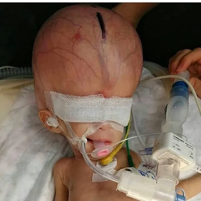
Hydrocephalus is cataloged in two ways. Depending on the origin, it can be congenital or acquired; and in relation to the CSF flow, this pathology is classified as communicating (i.e. CSF can exit the ventricular system, but the fluid is not absorbed readily, or there is too much fluid to be absorbed) or non-communicating (i.e. CSF cannot exit the ventricular system, and thus there is obstruction to CSF absorption, resulting in an increased pressure inside the brain). Symptoms of hydrocephalus may include an unusually large head with thin, transparent scalp, bulging forehead with increased spaces between the bones of the skull (fontanelles), and a downward gaze.
The most common congenital cause is the obstruction or narrowing (stenosis) of the cerebral aqueduct — the long, narrow passageway between the third and fourth ventricles of the brain. It can also be caused by infections, hemorrhage or a tumor.
The condition is pretty rare and can be easily detected early in pregnancy by ultrasound which does not necessarily shows the obstruction but detects an abnormal collection of fluid.
These kids are treated after birth by either inserting a shunt, small flexible plastic tube that diverts excess CSF from the brain to another part of the body where the fluid can be reabsorbed; or by an endoscopic ventriculostomy, where a small hole is made in the floor of the third ventricle, allowing the CSF to bypass the obstruction and flow toward other areas where reabsorption can happen.
Early medical illustrators like Andreas Vesalius understood this perfectly. They didn't just draw muscles and bones, they depicted "flayed" figures in dramatic, lifelike poses, emphasizing the humanity within the physical form. Today, modern imaging techniques like MRI and CT scans have replaced ink and parchment, but the reaction remains the same.
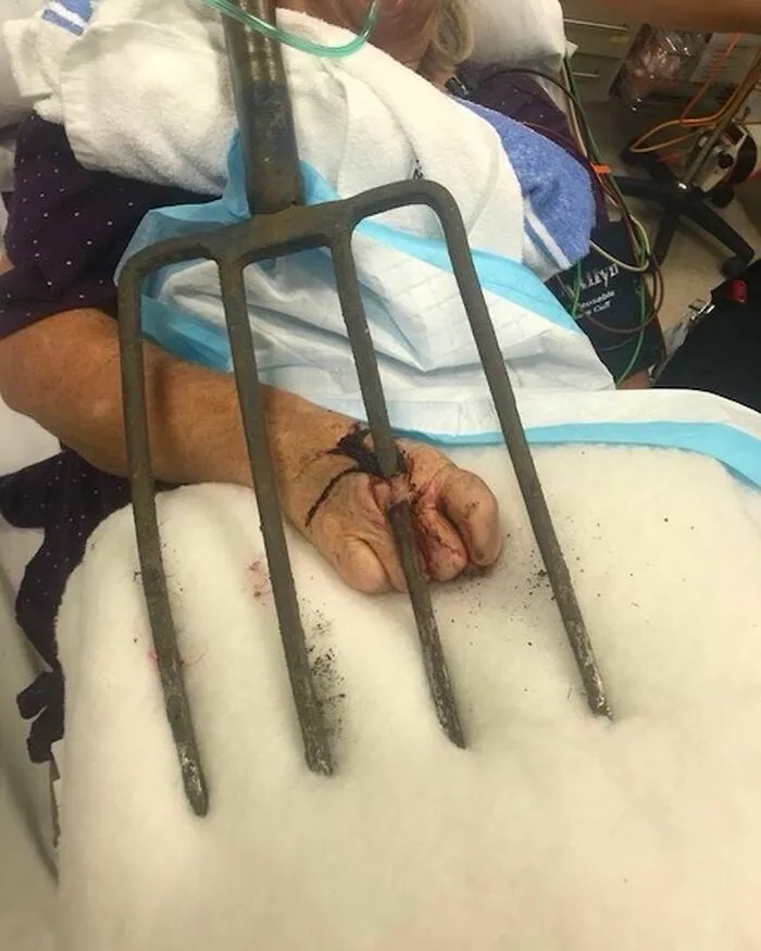
#22 What Started As A Peaceful Gardening Session Among The Blooms Quickly Became A Thorny Situation For This Nanny, As She Went To Hang Her Gardening Fork Up, It Fell And Took An Unintended Detour Into Her Hand
#23 This Man Miraculously Survived Being Impaled By A 40-Pound Piece Of Farm Equipment
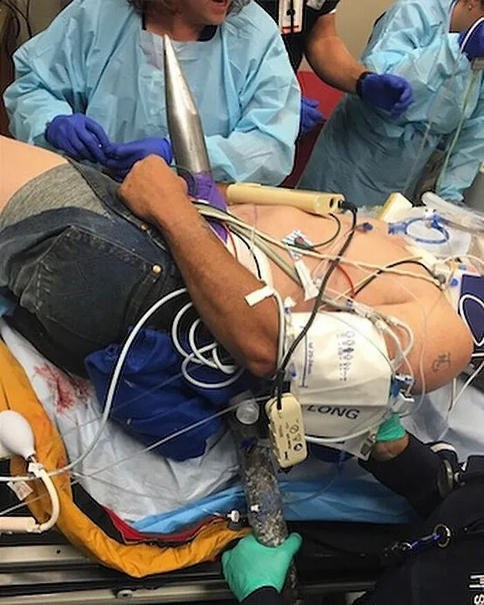
“I felt something hit me and I went to the ground, I looked up and I was looking around to find out what had happened, and I saw that belt tine sticking through me and into the ground.”
Since the spear, which weighed 40 pounds, was still attached to the machine when it went through his body, his coworkers had to cut it off from the loader with a heat torch.
He was flown to a medical centre and underwent a four-hour operation to remove the spear and repair internal injuries.
The object missed his spinal cord, his aorta and it missed the ureter — the tube connecting his kidney to his bladder by a few centimeters.
The patient was discharged only 2 weeks after the accident and is expected to make a full recovery.
While you would expect an accident like this to cause immediate death, some lucky people have been known to survive. If the object happens to miss vital organs, arteries or your spinal cord, doctors may be able to remove it and repair internal damage quickly enough to keep the patient alive.”
He did make an extraordinary recovery.
#24 56 Year Old Patient With Severe Toe Misalignment Due To Rheumatoid Arthritis, Before And After Surgery
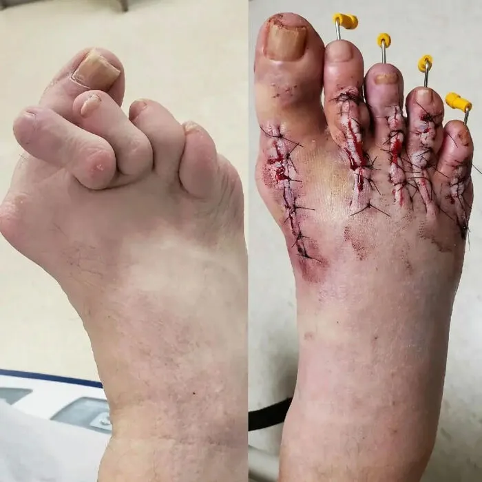
Rheumatoid arthritis is a chronic disease that attacks multiple joints throughout the body. It most often starts in the small joints of the hands and feet, and usually affects the same joints on both sides of the body.
More than 90% of people with rheumatoid arthritis (RA) develop symptoms in the foot and ankle over the course of the disease.
The joints of your body are covered with a lining — called synovium — that lubricates the joint and makes it easier to move. Rheumatoid arthritis causes an overactivity of this lining. It swells and becomes inflamed, destroying the joint, as well as the ligaments and other tissues that support it. Weakened ligaments can cause joint deformities — such as claw toe or hammer toe. Softening of the bone (osteopenia) can result in stress fractures and collapse of bone.
Fusion of the affected joints is the most common type of surgery performed for RA. Fusion takes the two bones that form a joint and fuses them together to make one bone.
During the surgery, the joints are exposed and the remaining cartilage is removed. The two bones are then held together with screws or a combination of screws and plates. This prevents the bones from moving.
Because the joint is no longer intact, this surgery does limit joint motion. Limited joint motion may not be felt by the patient, depending on the joints fused. The midfoot joints often do not have much motion to begin with, and fusing them does not create increased stiffness.
We are looking at a map of ourselves, but it’s a map written in a language we barely understand. The "creepiness" comes from the realization that there is a whole world functioning inside us, complete with its own wars, waste management systems, and repairs, that we have zero conscious control over.
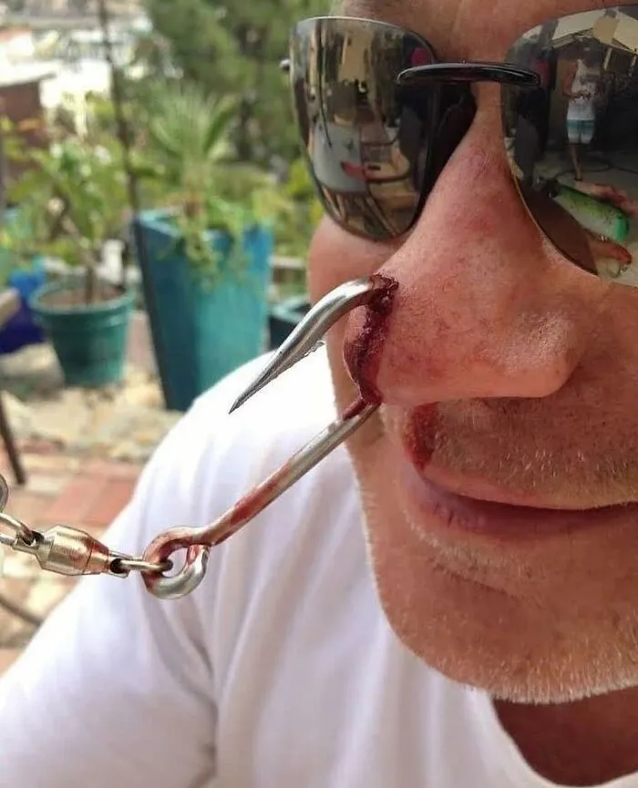
#26 These X-Ray Images Show Severe Skull And Facial Injuries Following A Gunshot From An AK-47
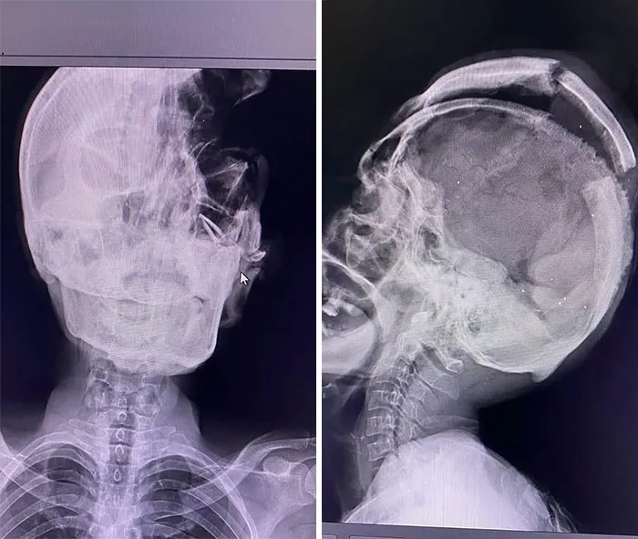
#27 A Rare Case Of Ovarian Ectopic Pregnancy
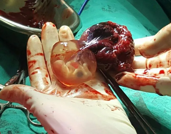
In other words, the fertilized egg implants and develops within the ovary instead of the uterus. It occurs when the fertilized egg fails to travel down the fallopian tube and instead implants in the ovarian tissue.
Clinically, it manifests with a constellation of symptoms including abdominal pain, frequently localized to one side, and vaginal bleeding. The symptomatology may demonstrate variable intensity, while concurrently mimicking other gynecological conditions, thereby precipitating diagnostic challenges and potential delays in appropriate management.
Diagnostic modalities encompass the utilization of transvaginal ultrasonography, which reveals the presence of a gestational sac within the ovarian milieu. Additional sonographic findings may encompass adnexal masses, augmented vascularity within the ovarian bed, or the absence of a gestational sac within the uterine cavity. Serial monitoring of serum human chorionic gonadotropin (hCG) levels contributes to the diagnostic algorithm, facilitating the assessment of pregnancy progression and viability.
Surgical intervention is frequently preferred and is typically accomplished through laparoscopic means, which confer enhanced visualization and precision in the removal of the ectopic pregnancy while minimizing collateral ovarian damage. However, instances necessitating extensive ovarian involvement or situations wherein laparoscopic proficiency is limited may necessitate recourse to laparotomy. The overarching goal of surgical intervention is the complete eradication of the ectopic pregnancy while preserving the functional integrity of the ovarian tissue.
Alternatively, selected cases may be amenable to medical management, wherein the administration of methotrexate, a folic acid antagonist, retards the growth of trophoblastic tissue, facilitating gradual resorption of the ectopic pregnancy by the body.
Furthermore, medical photography often highlights the fragility of the human condition, which is perhaps the most horrifying realization of all. Seeing a skull with a unique deformity or a heart enlarged by a lifelong struggle reminds us of the precariousness of health. It strips away the social masks we wear and reduces us to our base components. Yet, in that reduction, there is also a sense of profound connection. Every person who has ever lived has carried this same complex machinery within them.
#28 This Is Why Many Peoples’ Nightmares Contain Spiders. An Extreme Case Showing Brown Recluse Spider Bite Of The Thumb

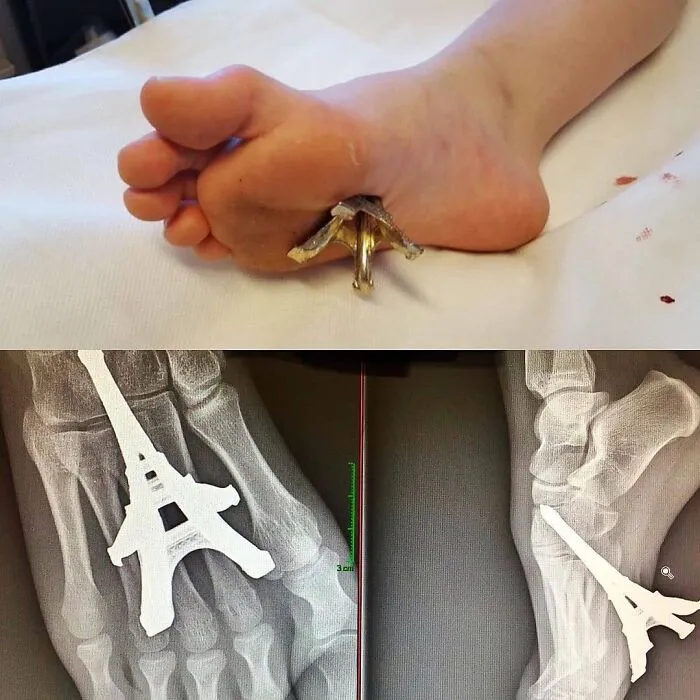
#29 “Ei-Ffel”. This Is The Actual Photo Of Yesterday’s Case Showing An Eiffel Tower Miniature Embedded Into The Foot
#30 Man With 3-Inch Nail In Skull Goes To Hospital After Two Days… But Can’t Remember How It Got There
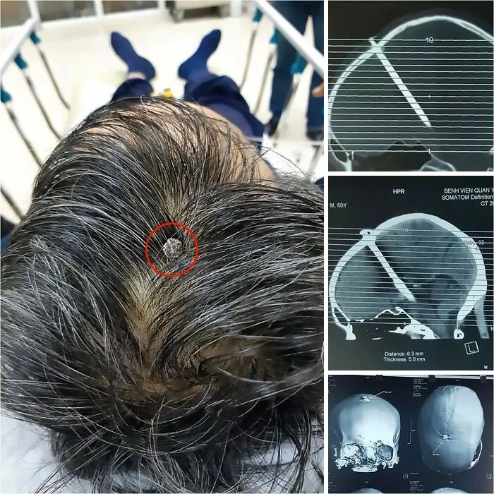
The Emergency Department staff soon found the reason for the man's headache; a large iron nail lodged in the patients head, which seems to have been hammered into the patient’s skull from the top down. OUCH!
The weird part is that he has no memory of how it ended up there.
He was completely conscious and didn’t have any traces of blood surrounding his wound.
He was rushed straight into a three-hour operation to remove the nail after revealing the depth of the injury in a CT scan.
By studying the "gross" or the "weird" aspects of medicine, we are actually engaging in a deep act of empathy and self-discovery. We are trying to understand the vessel that carries our consciousness through the world, even if that vessel looks a bit terrifying when you take the lid off. Ultimately, we stare because we cannot look away from the miracle of our own survival, even when that miracle is wrapped in something that makes our skin crawl.
#31 The Human Body Is Truly Amazing. This Photo Shows You A Rare Glimpse At Something That’s Called Fascia, It’s The White Covering That Is On Top Of Muscle And Under The Skin
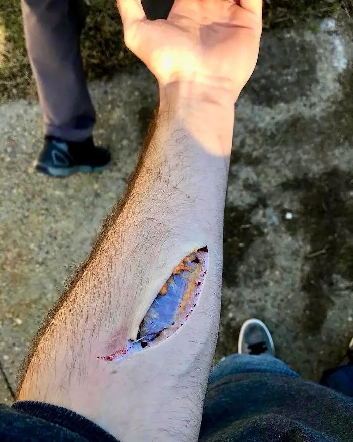
Fascia can be broken down into four main layers: superficial, deep, visceral, and parietal.
Superficial fascia is right under the skin. It’s thicker in the main part of the body (stomach, chest, etc.), and it gets thinner in places further away, like in the hands and feet.
Deep fascia covers bones, muscles, nerves, and blood vessels.
The visceral fascia layer goes around certain organs that settle into the body’s open spaces, including the lungs, heart, and stomach.
The injury in this case was caused by a screw bolt that was sticking out of a stove! The patient was injured when he pushed a stove off of a dolly that was about to fall on a coworker. He saved the coworker from harm, but unfortunately was cut on the arm in the process by a bolt sticking out of the stove. Resulting in the injury shown in the photos. The last photo shows the scar healed one year after the injury.
#32 Ring Got In Contact With A Car Battery, Causing Circumferential Burn To The Fourth Finger!
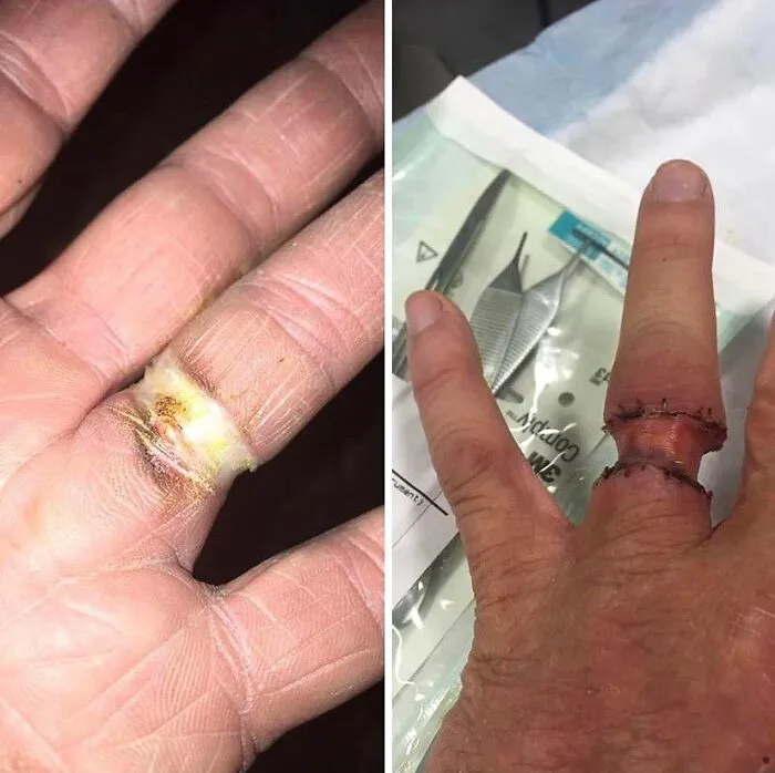
Electrical burns associated with rings are usually deep second-degree burns which may leave scars, cause loss of hair follicles and sweat glands, leave permanent skin pigmentation and may need skin grafting. The outcome depends on the severity of damage to the underlying tissue especially digital blood supply.
#34 Laser Tattoo Removal Gone Wrong — Huge Blisters Containing Ink While Removing Tattoos
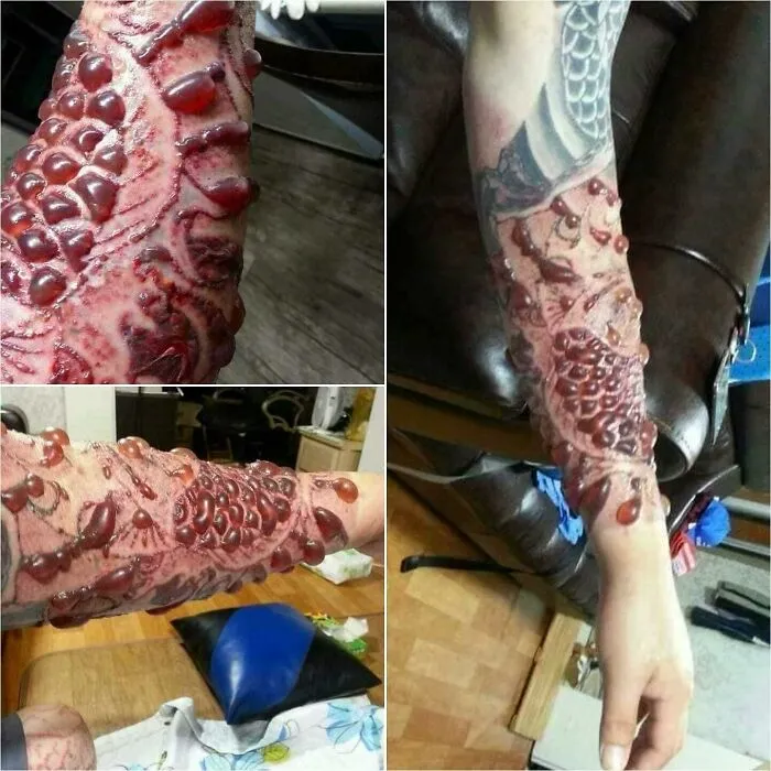
In laser tattoo removal, the laser emits high-energy pulses targeted at the tattoo ink, causing it to fragment into smaller particles that the body can eliminate. The intense heat generated (thousands of degrees) during this process may result in thermal injury to the surrounding skin, prompting an inflammatory response. Blisters may form as a consequence of increased permeability and fluid accumulation in the epidermal and dermal layers.
Essentially the blisters are containing ink, blood and water as the laser heating up the ink particles in the skin which can then cause blood vessels surrounding the tattoo to rupture.
Proper post-procedural care, including wound management and infection prevention, is essential to support optimal healing and reduce the risk of complications.
#35 Putting That Skull Back In, Just Like A Puzzle!
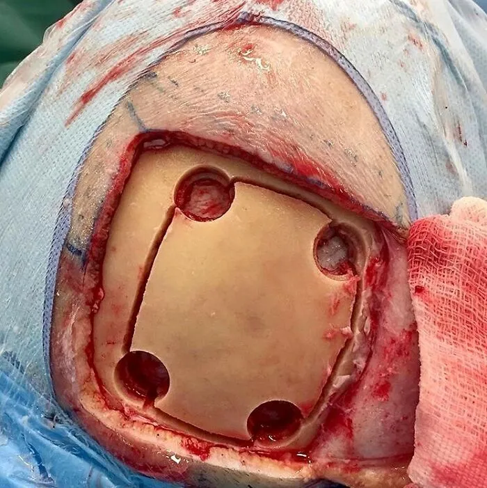
Craniotomy involves the removal of a segment of the cranium to gain access to the intracranial contents for brain surgery.
After a scalp incision is made, a burr hole is created using a high-speed drill. This allows for the removal of a bone flap, which is carefully preserved.
Following the bone flap removal, the dura mater is typically incised, providing access to the brain. Various surgical techniques are employed, depending on the specific pathological condition being addressed. These may include tumor resection, hematoma evacuation, arteriovenous malformation excision, or the placement of intracranial monitoring devices.
After the necessary intervention is completed, the dura mater is sutured or sealed using synthetic materials. The bone flap is then repositioned and secured using titanium plates, sutures, or wire mesh. The bone will eventually re-grow and the skull will be fully continuous again.
In certain cases, when the bone flap cannot be reattached, alternative synthetic materials (like a titanium mesh) may be used to reconstruct the cranial defect and replace the bone flap if it cannot be reattached.
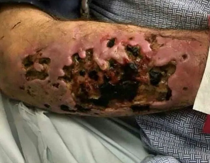
#36 A Drug With A Nasty Bite, Here’s A Close-Up Of A Krokodil-Induced Skin Necrosis In An Iv Drug User
#37 A Dodgeball Injury That Resulted In Radius And Ulnar Shaft Fracture, AKA Broken Forearm
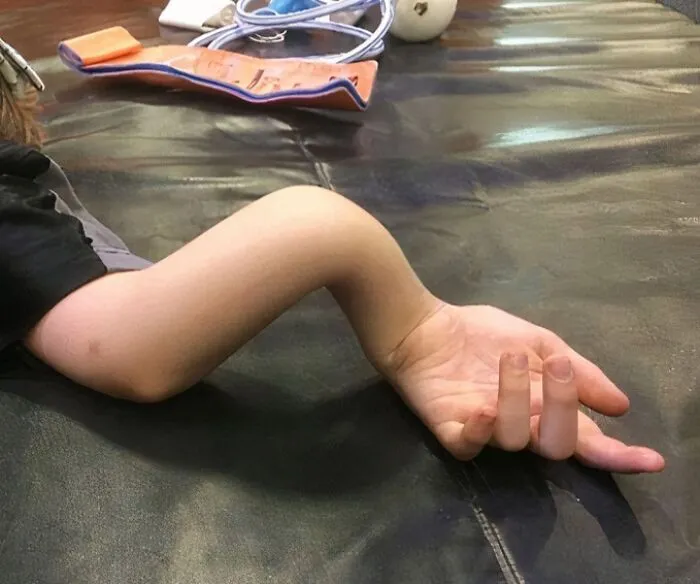
This particular injury entails the disruptive discontinuity of the long bones situated within the antebrachial region, namely the radius and ulnar diaphyses. Etiologically, such fractures arise consequent to an abrupt forceful impact or trauma, commonly encountered within dynamic sports such as dodgeball. Timely medical intervention is imperative to accurately ascertain the nature and extent of this injury, often accomplished through radiographic imaging modalities like X-ray examinations. Treatment modalities encompass immobilization employing a cast or, in severe instances, surgical intervention to achieve anatomical alignment and biomechanical stability of the fractured bony elements.
Due to significant displacement, the initial treatment is closed reduction that is attempted when fracture angulation exceeds 10 degrees or displacement exceeds 50%.
Reduction involves placing the patient's affected arm in finger traps while the brachium is secured with a strap or weights. This setup allows for ready manipulation of the forearm and rapid splinting once reduction is achieved. After satisfactory alignment is achieved, the fractured arm is placed in a long-arm posterior splint with the elbow at 90 degrees and the wrist in neutral (ie, without supination or pronation) and slight extension.
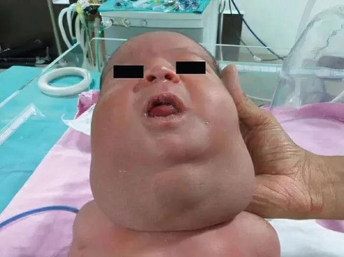
#38 Cystic Hygroma It Is A Fluid-Filled Sac Due To Blockage Of The Lymphatic System That Appears As A Swelling Usually In The Neck
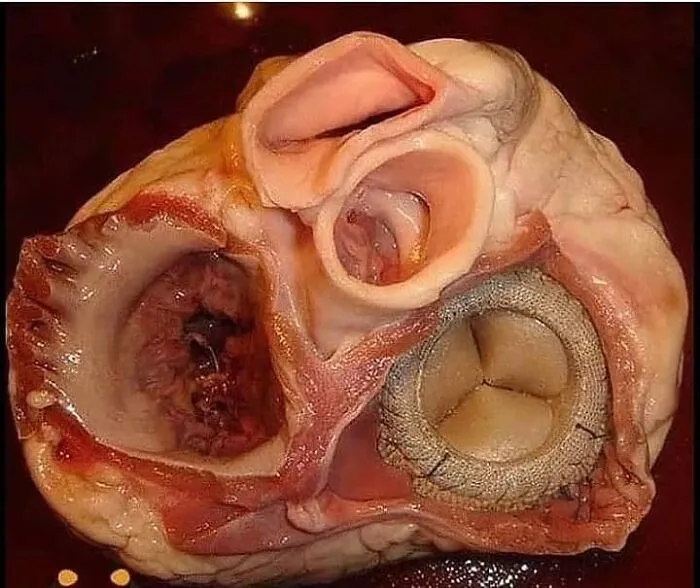
#40 Cross Section Of The Lungs Of A Patient With Copd (Emphysema , Chronic Bronchitis And Asthma)

