







Do you know what the universal blood type is? Apparently, only one in 10 Americans do. (It’s O-negative, by the way.) How much do you know about the lymphatic system? And do you know if you’re getting enough dietary fiber every day? (Chances are, you aren’t.) The reality is that the human body is a mystery to the majority of us. And even if you’ve been to medical school, there might be plenty of conditions you’ve never encountered.
To learn more about what’s going on in the mysterious world of medicine, we took a trip to Medical Doctors on Instagram. This page shares a variety of photos and information to educate others about rare conditions and provide a glimpse into what's going on inside our hospitals. We’ll warn you right now that some of these images are definitely not for pandas with weak stomachs, and other photos might break your heart. But if you can make it through this list, we hope you’ll learn something new!
#42
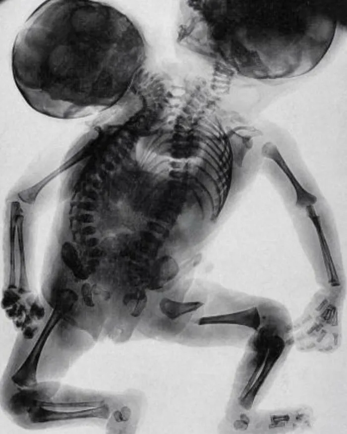
An X-ray of conjoined twins born with two backbones and two heads
Conjoined twins are two babies who are born physically connected to each other. Conjoined twins develop when an early embryo only partially separates to form two individuals. Although two babies develop from this embryo, they remain physically connected — most often at the chest, abdomen or pelvis. Conjoined twins may also share one or more internal body organs. Though many conjoined twins are not alive when born (stillborn) or die shortly after birth, advances in surgery and technology have improved survival rates. Some surviving conjoined twins can be surgically separated. The success of surgery depends on where the twins are joined and how many and which organs are shared. It also depends on the experience and skill of the surgical team.
Report
14points
#43
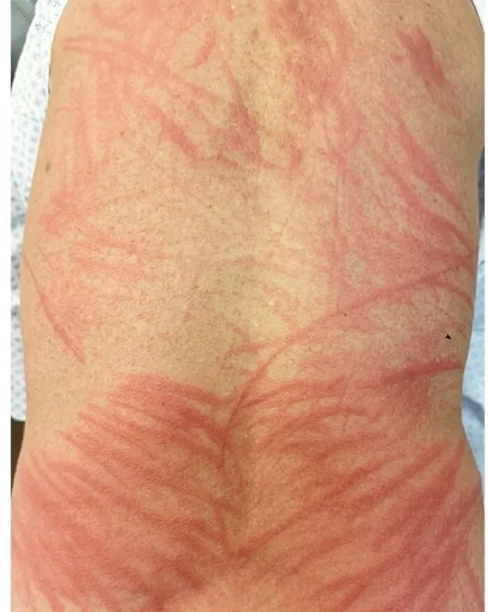
A 72-year-old man presented to the emergency department with a 2-day history of an itchy rash on his back. On physical examination, edematous, flagellate plaques and linear patches were present across the patient's entire back and upper buttocks. There was no adenopathy, dermographism, or mucosal involvement
Report
13points
#44
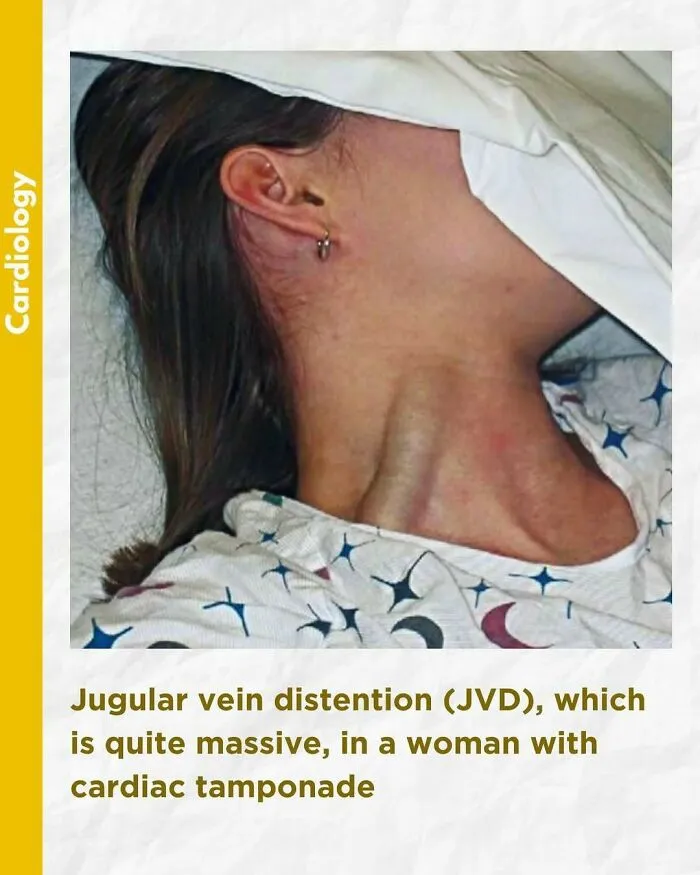
An elevated jugular venous pressure (JVP) is the classic sign of venous hypertension (e.g. right-sided heart failure). JVP elevation can be visualized as jugular venous distension, whereby the JVP is visualized at a level of the neck that is higher than normal. The jugular venous pressure is often used to assess the central venous pressure in the absence of invasive measurements (e.g. with a central venous catheter, which is a tube inserted in the neck veins).
Report
13points
#45
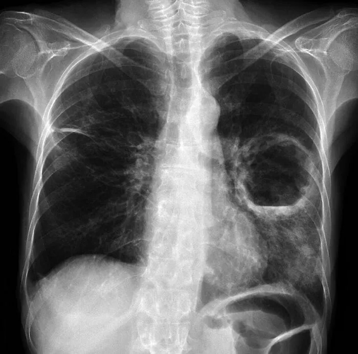
Chest x-ray of a patient demonstrates a large left sided pulmonary cavity with a small dependent air-fluid level within the left mid zone, in keeping with pulmonary abscess. Patchy airspace opacification more inferiorly within left lower zone.
Scarring/atelectasis within lateral aspect of right upper zone.
Scarring/atelectasis within lateral aspect of right upper zone.
Report
12points
#46
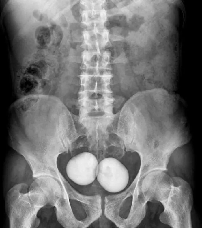
KUB X-ray shows two large and oval radio-opaque shadows in the pelvis
Report
12points
#47
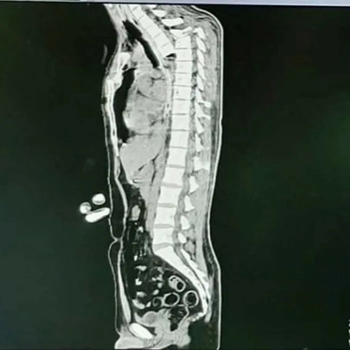
Presented is a case of spinal cord transection, where the spinal cord is cut in half. The spinal cord is the main relay station for both afferent sensory information and efferent motor commands. There are multiple sensory tracts which ascend the spinal cord such as the Dorsal Column-Medial Lemniscus (DCML) and spinothalamic. The DCML carries ipsilateral information of pressure, vibration, fine touch and proprioception from sensory nerve endings in the periphery, whereas the spinothalamic tract carries contralateral information of pain, temperature, pressure and crude touch. The main motor pathway that controls voluntary movement is the lateral corticospinal tract, although there are many others. In cases of spinal cord injury, the spinal cord often goes into spinal shock, which is a characteristic reflexive pattern of areflexia->hyporeflexia->hyperreflexia. The state of a patients reflexes can often be tested clinically by testing a reflex arc such as the patellar reflex. Unfortunately, the patient suffered massive spinal cord trauma, sternal fractures, with bilateral lung contusions leading to a hematothorax and expired
Report
11points
#48
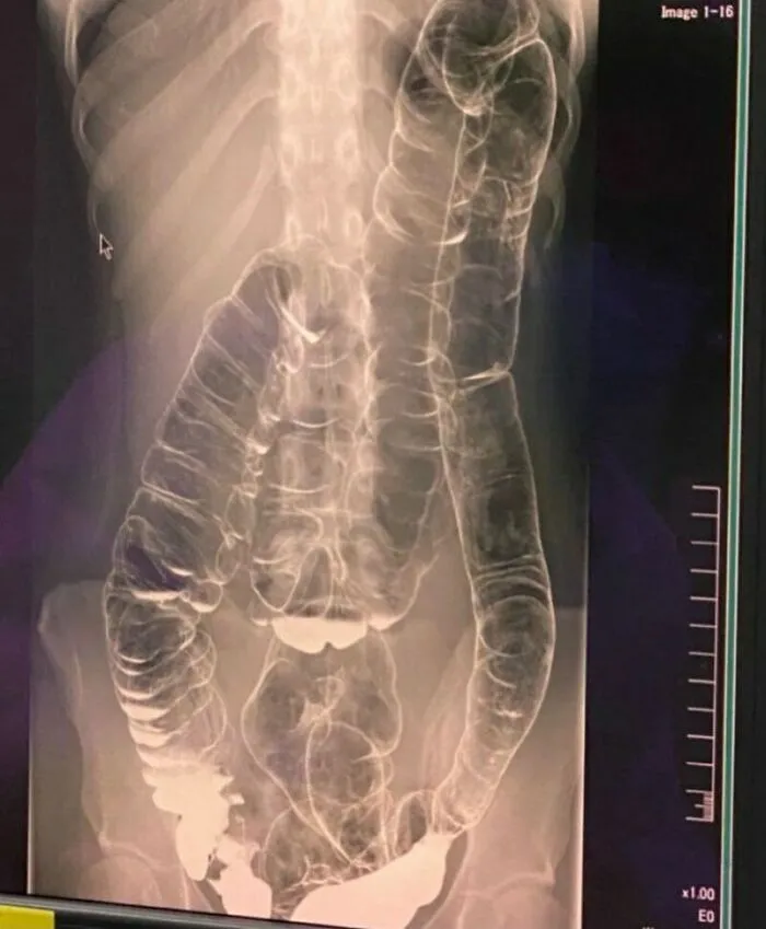
This is what constipation after 19 days looks like. A woman suffering from chronic constipation, discovers her colon severely displaced.
Report
11points
#49
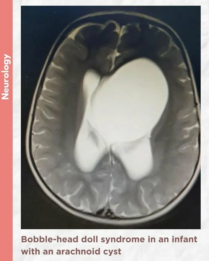
Bobble-head doll syndrome in an infant with an arachnoid cyst
Bobble-head doll syndrome is a rare neurological movement disorder in which patients, usually children around age 3, begin to bob their head and shoulders forward and back, or sometimes side-to-side, involuntarily, in a manner reminiscent of a bobble head doll. The syndrome is related to cystic lesions and swelling of the third ventricle in the brain. Symptoms of bobble-head doll syndrome are diverse but can be grouped into two categories: physical and neurological. The most common form of treatment is surgical implanting of a shunt to relieve the swelling of the brain.
A 1.5-year-old girl presented to the pediatric clinic with the chief complaints of gradual onset excessive head nodding (side-to-side movement) for 3 months. Movements increased with walking, emotions, and stress; decreased during periods of concentration; and were absent during sleep. There were no other complaints or headaches. There was no other significant history.
The child was alert, with normal cognitive function. Neurological examination was normal. Initial laboratory assessment including CBC, hepatic and renal function, and endocrine function tests were normal.
Cranial MRI demonstrated a large left-hemispheric cystic process with a midline shift, well-defined thin-walled suprasellar arachnoid cyst measuring 3 × 5 × 7 cm that obstructed the foramina of Monro, with resulting hydrocephalus ventriculomegaly. Based on the cranial MRI and symptoms, a diagnosis of a suprasellar arachnoid cyst with BHDS was made. The patient underwent endoscopic cystoventriculostomy and cystocisternostomy for the suprasellar arachnoid cyst. During the 6 months of follow-up, the head bobbing disappeared completely, and her growth was normal.
Despite the rareness of bobble-head doll syndrome, it is considered an important condition that must be investigated early to detect the cause and treated promptly to avoid potential complications.
A 1.5-year-old girl presented to the pediatric clinic with the chief complaints of gradual onset excessive head nodding (side-to-side movement) for 3 months. Movements increased with walking, emotions, and stress; decreased during periods of concentration; and were absent during sleep. There were no other complaints or headaches. There was no other significant history.
The child was alert, with normal cognitive function. Neurological examination was normal. Initial laboratory assessment including CBC, hepatic and renal function, and endocrine function tests were normal.
Cranial MRI demonstrated a large left-hemispheric cystic process with a midline shift, well-defined thin-walled suprasellar arachnoid cyst measuring 3 × 5 × 7 cm that obstructed the foramina of Monro, with resulting hydrocephalus ventriculomegaly. Based on the cranial MRI and symptoms, a diagnosis of a suprasellar arachnoid cyst with BHDS was made. The patient underwent endoscopic cystoventriculostomy and cystocisternostomy for the suprasellar arachnoid cyst. During the 6 months of follow-up, the head bobbing disappeared completely, and her growth was normal.
Despite the rareness of bobble-head doll syndrome, it is considered an important condition that must be investigated early to detect the cause and treated promptly to avoid potential complications.
Report
11points
#50
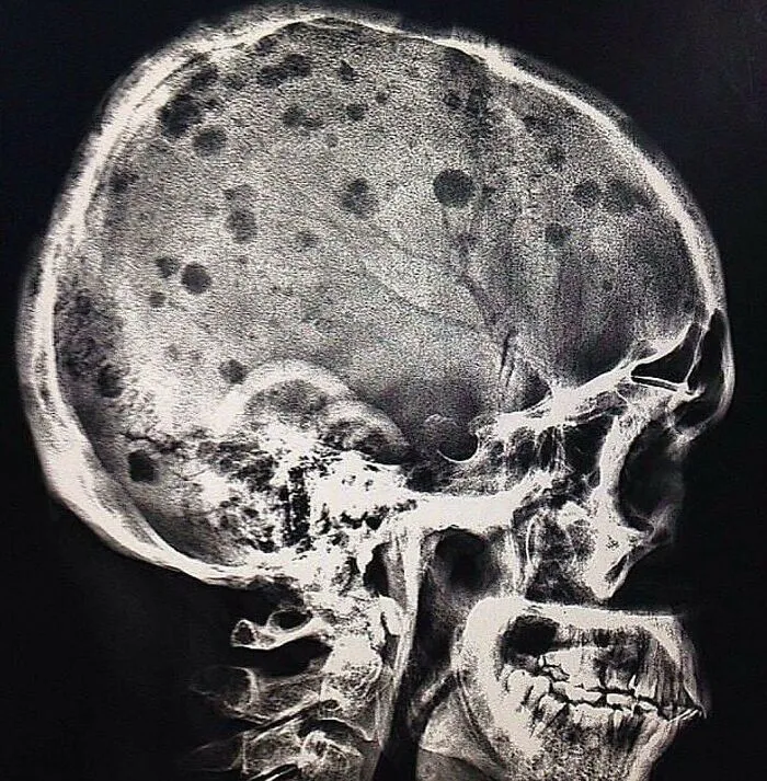
Lateral view skull radiograph of a patient with multiple myeloma
The classic radiographic appearance of multiple myeloma is that of multiple, small, well-circumscribed, lytic, punched-out, round lesions within the skull, spine, and pelvis. The pattern of lytic or punched-out radiolucent lesions on the skull have been described as resembling raindrops hitting a surface and splashing. How would you manage a patient with multiple myeloma?
Report
9points
