



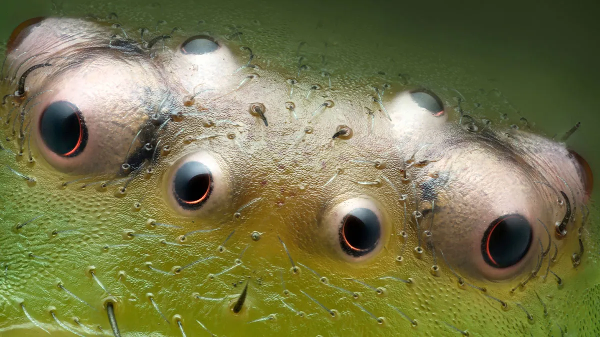


The 2024 Nikon Small World Photomicrography Competition, now in its 50th year, celebrates the beauty and science behind the smallest details of our world. Each year, scientists and artists from around the globe submit stunning microscope images that reveal extraordinary views of life on a microscopic scale. From intricate cell structures to fascinating natural phenomena, these images offer a unique glimpse into the hidden world around us.
This year’s winners did not disappoint. First place was awarded to Dr. Bruno Cisterna for his incredible image of mouse brain tumor cells, which sheds light on neurodegenerative diseases like Alzheimer's and ALS. The 2024 competition continues to highlight how microscopy advances both art and science.
#21 9th Place - John-Oliver Dum

Medienbunker Produktion
Bendorf, Rheinland Pfalz, Germany
Bendorf, Rheinland Pfalz, Germany
"Pollen in a garden spider (Araneus) web."
Report
38points
#22 Image Of Distinction - Dr. Saikat Ghosh

National Institutes of Health
NICHD
Bethesda, Maryland, USA
NICHD
Bethesda, Maryland, USA
"Human neurons."
Report
38points
#23 Image Of Distinction - Joshua Coogler

Dallas, North Carolina, USA
"Moss sporophyte with spores (green)."
Report
37points
#24 Image Of Distinction - Daniel Knop

Oberzent-Airlenbach, Hessen, Germany
"Opening of a hibiscus flower (Hibiscus moscheutos) exposing the pollen in four stages, each ten minutes apart."
Report
37points
#25 11th Place - Dr. Ferenc Halmos

Bánd, Veszprém, Hungary
"Slime mold on a rotten twig with water droplets."
Report
36points
#26 Image Of Distinction - Daniel Knop

Oberzent-Airlenbach, Hessen, Germany
"Dorsal part of cuckoo wasp (Hedychrum gerstaeckeri) abdomen."
Report
36points
#27 Image Of Distinction - Jacek Myslowski

Wloclawek, Kujawko-Pomorskie, Poland
"Water mite (Arrenurus)."
Report
34points
#28 16th Place - Marek Miś

Suwalki, Podlaskie, Poland
"Two water fleas (Daphnia sp.) with embryos (left) and eggs (right)."
Report
33points
#29 Image Of Distinction - Dr. Igor Robert Siwanowicz

Howard Hughes Medical Institute (HHMI), Janelia Research Campus
Ashburn, Virginia, USA
Ashburn, Virginia, USA
"Aster anther cross-section with pollen grains (green)."
Report
33points
#30 Image Of Distinction - Dr. Robert Markus

University of Nottingham
School of Life Sciences, Super Resolution Microscopy
Nottingham, Nottinghamshire, United Kingdom
School of Life Sciences, Super Resolution Microscopy
Nottingham, Nottinghamshire, United Kingdom
"Dandelion (Traxacum officinale) cross section showing curved stigma with pollen."
Report
32points
Understanding the raw and gritty struggles faced by communities is crucial, much like unveiling intricate details only visible under a microscope. At Kensington Avenue, residents share their poignant stories which reflect societal issues at their core, akin to the way photomicrography opens up hidden worlds of science.
Delve deeper into these revelations about life’s unseen facets by exploring the detailed documentation of raw experiences on the challenges faced by a particular Philadelphia community.


