



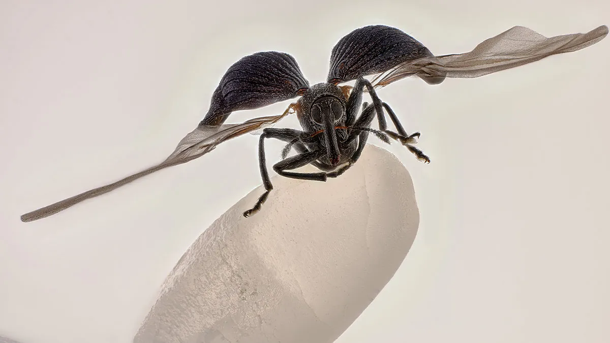


The winners of the 51st annual Nikon Small World Photomicrography Competition have been announced, showcasing some of the most stunning and detailed images of the microscopic world. From insects to plant structures, these photographs reveal the beauty and complexity of tiny subjects we often overlook in our daily lives. The competition, run by Nikon Instruments, has been celebrating excellence in microscopy and digital imaging for over five decades, inspiring scientists and artists alike.
This year’s top prize went to China’s Zhang You for his incredible image of a rice weevil perched on a grain of rice, wings fully extended. Using a combination of photography skills, careful lighting, and focus stacking, You captured a moment that blends scientific detail with artistic flair. His work reminds us that even the smallest creatures can be fascinating and beautiful when seen up close.
#41 Image Of Distinction: Daniel Han, Diatoms Australia (Macro Cosmos Imaging), Sydney, New South Wales, Australia
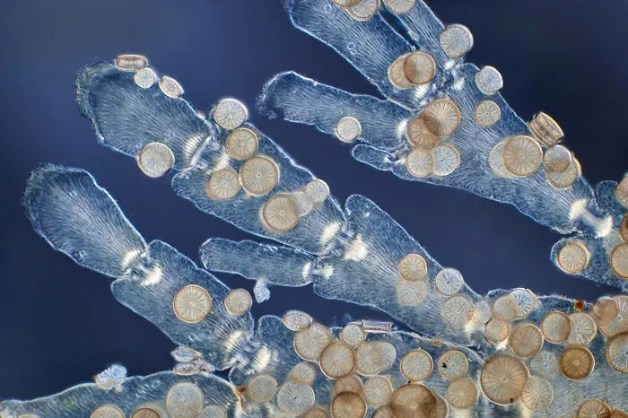
"Diatoms (Arachnoidiscus sp.) on coralline algae."
5points
#42 Image Of Distinction: Doong Yien, Xmato Works, Beijing, China
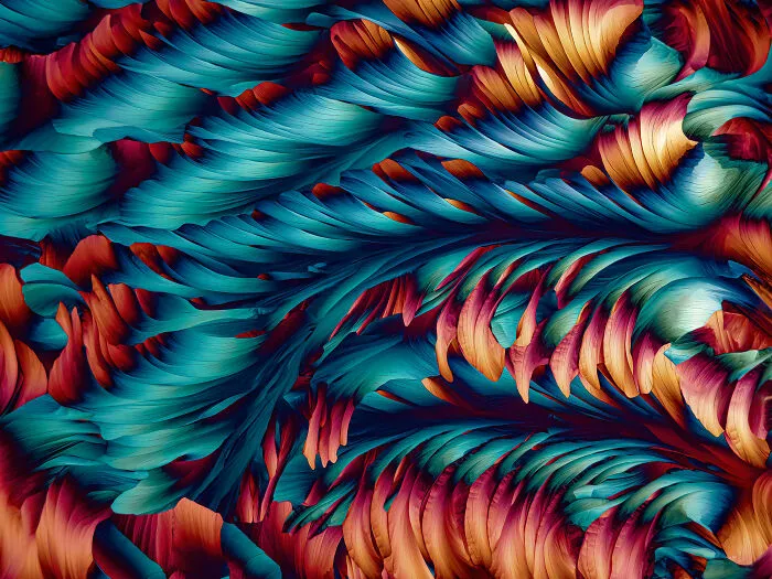
"Crystallization of a mixed solution of alanine and glutamine under polarized light."
5points
#43 Image Of Distinction: Hannah Somers, Mdi Biological Laboratory, Light Microscopy Facility, Bar Harbor, Maine, USA
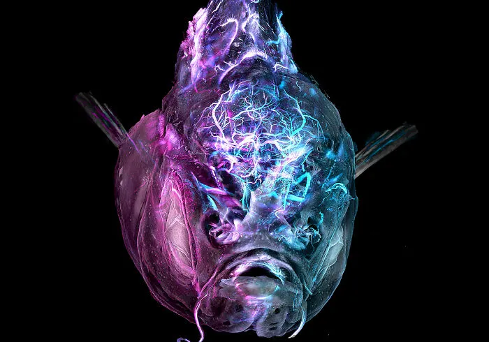
"An adult zebrafish showing blood vessels in the brain."
5points
#44 Image Of Distinction: Heiti Paves, Tallinn, Harju, Estonia
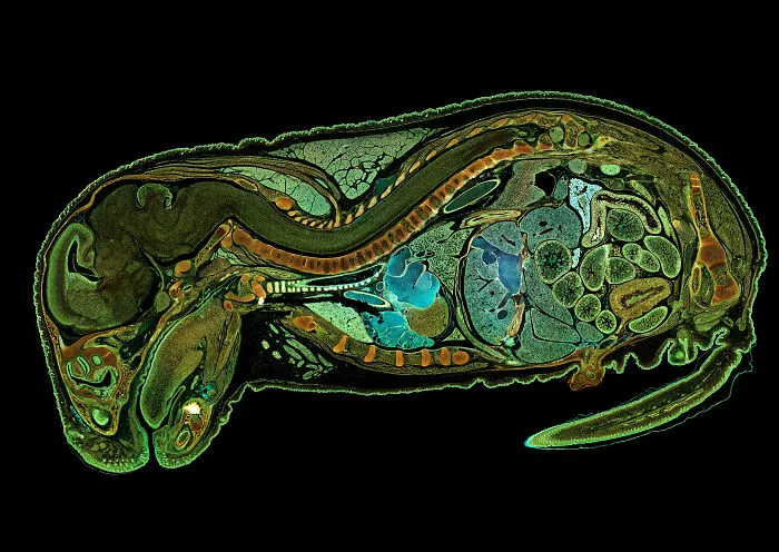
"Mouse embryo, sagittal section."
5points
#45 Image Of Distinction: Dr. Julien Resseguier, University Of Oslo, Department Of Biosciences / Fyscell, Oslo, Viken, Norway
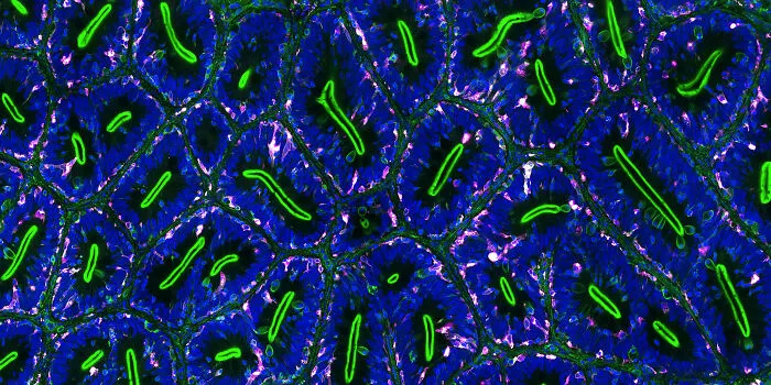
"Immune cells (magenta) protecting the different tissue compartments of the zebrafish intestines."
5points
#46 Image Of Distinction: Shambhavi Dwivedi And Dr. Friedemann Kiefer, University Of Münster, Faculty Of Biology, Muenster, North-Rhine Westphalia, Germany
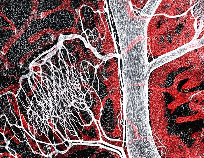
"Mouse lymphatic network (red) flanking blood vessels (white)."
5points
#47 Honorable Mention: Rebecca Lee, Yale University, Department Of Genetrics, New Haven, Connecticut, USA
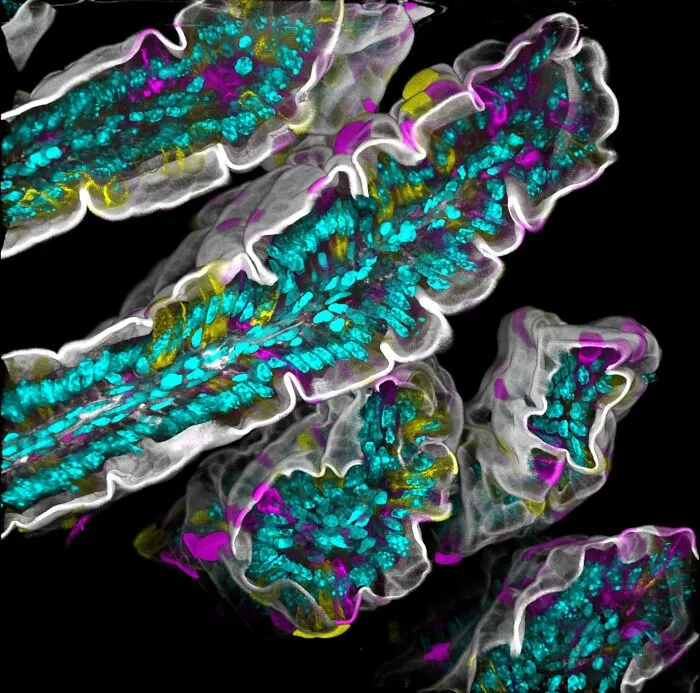
"Villi in the mouse small intestine."
4points
#48 Image Of Distinction: Dr. Rory L. Cooper And Professor Michel Milinkovitch, University Of Geneva, Department Of Genetics And Evolution, Geneva, Switzerland
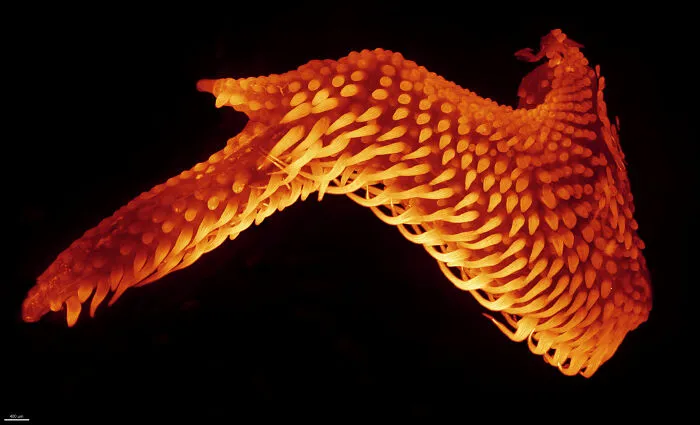
"Wing of the chicken embryo after 11 days of development."
4points
#49 10th Place: Dr. Dylan Burnette And Dr. James Hayes, Vanderbilt University School Of Medicine, Department Of Cell And Developmental Biology, Nashville, Tennessee, USA
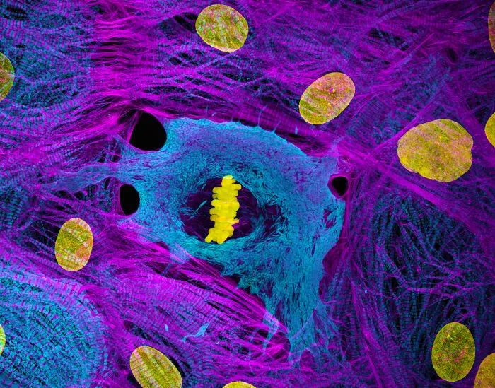
"Heart muscle cells (iPSC-derived) showing condensed chromosomes in metaphase."
4points
#50 4th Place: Dr. James Hayes, Vanderbilt University, Department Of Cell And Developmental Biology, Nashville, Tennessee, USA
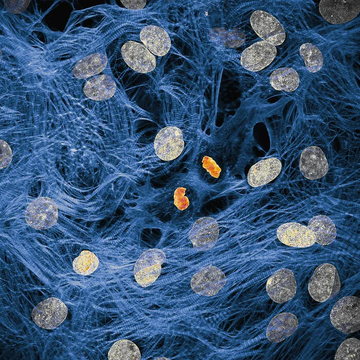
"Heart muscle cells with chromosomes condensed following cell division."
3points


